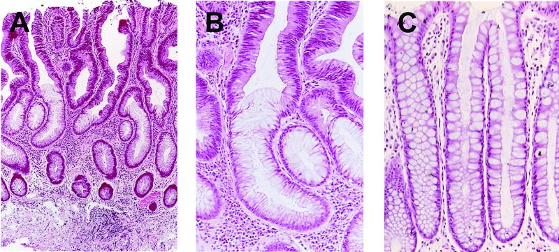
Figure 1.
(A) Hematoxylin and eosin stained section of a typical small adenomatous polyp. Dysplastic epithelium is located superficially in the crypts and is contiguous with the underlying histologically normal epithelium at the base. (B) Note the abrupt transition between dysplastic and normal-appearing epithelial cells at the mid-point of this crypt. (C) Adjacent normal colonic mucosae for comparison.

