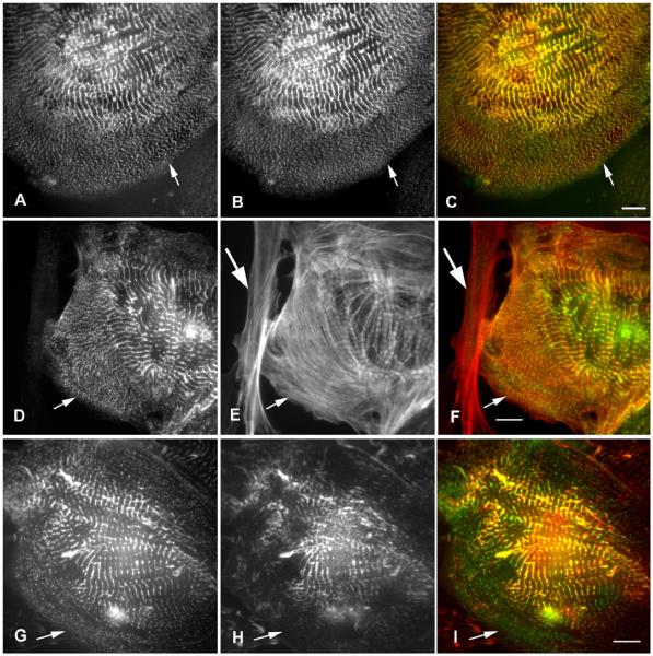
Figure 4.
(A-I) Co-staining of cardiomyocytes with anti-ArgBP2 antibody (A, D, G) and anti-sarcomeric alpha-actinin antibody (B); phalloidin (E); or anti-vincluin antibody (H). (A-C) ArgBP2 (A, green in C) co-localizes with alpha-actinin (B, red in C) in the Z-bodies (arrows) in the spreading lamella of a cardiomyocyte where myofibrils are known to assemble (Dabiri et al., 1997) and in Z-bands in the center of the cell. (D-F) ArgBP2 (D, green in F) localizes in the Z-bands in the middle of the phalloidin-stained I bands and in a beaded distribution along the unbanded phalloidin-stained actin fibers (E, red in F) in the spreading edge (small arrows) of the cardiomyocyte. ArgBP2 does not localize in actin fibers in the fibroblast on the left side of the image (E, F large arrow). (G-I) ArgBP2 (G, green in I) colocalizes with concentrations of vinculin (H, red in I). In addition ArgBP2 localizes in Z-bodies (arrows, H, I) and some Z-bands where vinculin is not present. Bars = 10 microns.

