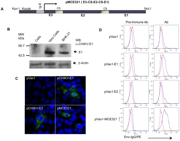
Figure 2. Construction and characterization of CHIKV DNA vaccine.
(A) Schematic representation of pMCE321 construct. The flanking enzyme sites used for cloning, Kozak expression element, CMV promoter, human IgE-leader, CHIKV fusion gene (E3-E2-E1), and cleavage sites (CS) are indicated and were cloned into the pVax1 vector. (B) Expression of pMCE321 constructs was confirmed in vitro using Envelope-E1 antiserum for the Western blot of CHIKV envelope antigens expressed in Vero and BHK-21 cells by Western blotting. Arrows indicate the positions of E1 protein expression. (C) Immunofluorescent assay showing staining of Vero cells transfected with pCHIKV-E1, pCHIKV-E2, or pMCE321 constructs and transient expression of the envelope proteins. (D). FACS analysis of envelope expression in transfected cells (0.5×10⁶ cells). Vero cells were transfected with indicated constructs and stained with anti-Env sera raised in mice, followed by staining with secondary PE-conjugated anti-mouse IgG antibody as indicated. Two representative FACS histograms are shown.

