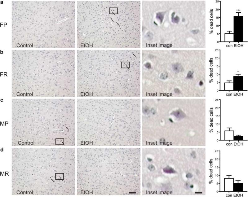
Figure 4.
Assessment of brain damage in male and female WSP and WSR following chronic EtOH exposure and withdrawal for 10 days. H&E staining was performed and arrows indicate eosinophilic staining characteristic of dead or dying cells (× 100 magnification). Zoomed regions (indicated by square inset region) showing morphology and staining of dead neurons from EtOH females or control males are included. (a) H&E stained sections from control or EtOH withdrawn female WSP mice. The percentage of damaged cells after EtOH withdrawal was significantly elevated in female WSP (FP; ***P<0.0003) mice. (b) Similarly in WSR females (FR), EtOH withdrawal resulted in increased cell death (*P<0.05). (c) In contrast to females, EtOH withdrawal in WSP male (MP) mice resulted in a non-significant decrease in dead or dying cells. (d) Likewise, analysis in male WSR (MR) mice revealed a modest decrease in cell death. Data are mean±SEM. Bar = 100 μm for photomicrographs; 14 μm for zoomed images.

