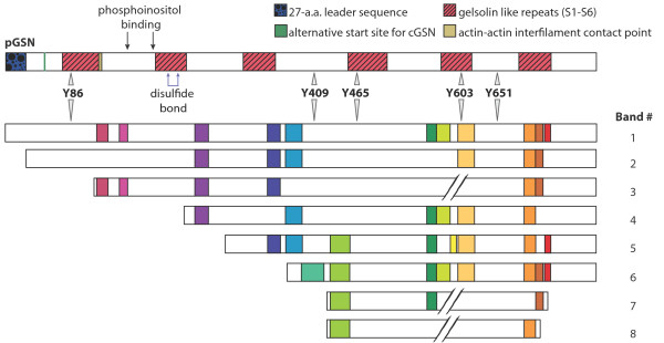
Figure 4.
Schematic model of full-length hGSN and proposed forms of gelsolin isolated from serum/plasma and CSF. Band 1 represents the full-length hGSN molecule and includes its functional and structural features. This form shows electrophoretic mobility corresponding to approximately 86 kDa. hGSN was identified by tandem mass spectrometry analysis in bands 2 to 8. Based on their electrophoretic mobility and identified peptides resulting from trypsin digestion (see Table 1 for details) we estimated their approximate molecular weight and amino acid sequence coverage. Gelsolin peptides identified in each band by LC/ESI-MS/MS are colored.

