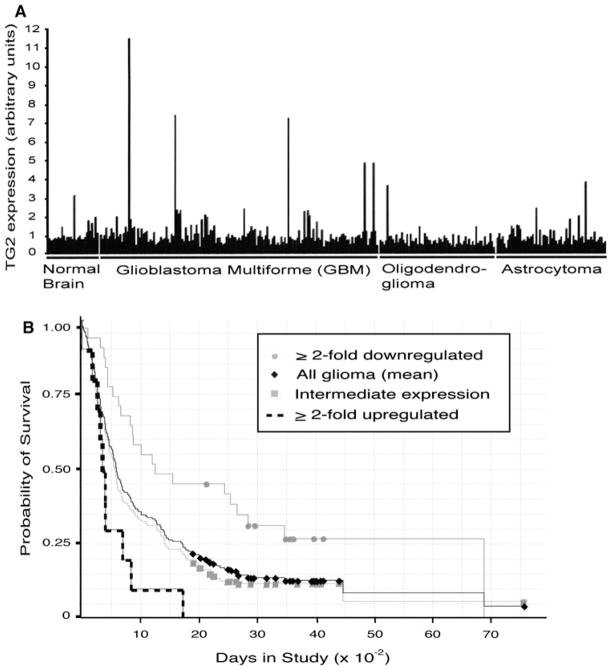
Fig. 4.
TG2 expression is heterogeneous in human brain tumors. a Relative TG2 expression on normal brain tissue, glioblastoma multiforme (stage IV), oligodendroglioma (stage II, III) or astrocytoma (stage II, III) specimens was obtained by query of the Rembrandt database. b Brain tumor samples were stratified into tumor groups that show ≥ 2-fold upregulation (dashed line), ≤ 2-fold downregulation (gray circles), and intermediate levels (gray squares) of TG2 expression. Kaplan–Meier analysis was subsequently conducted with these groupings, as well as mean TG2 expression among all glioma samples (black diamonds), to compare the association of TG2 expression with patient survival

