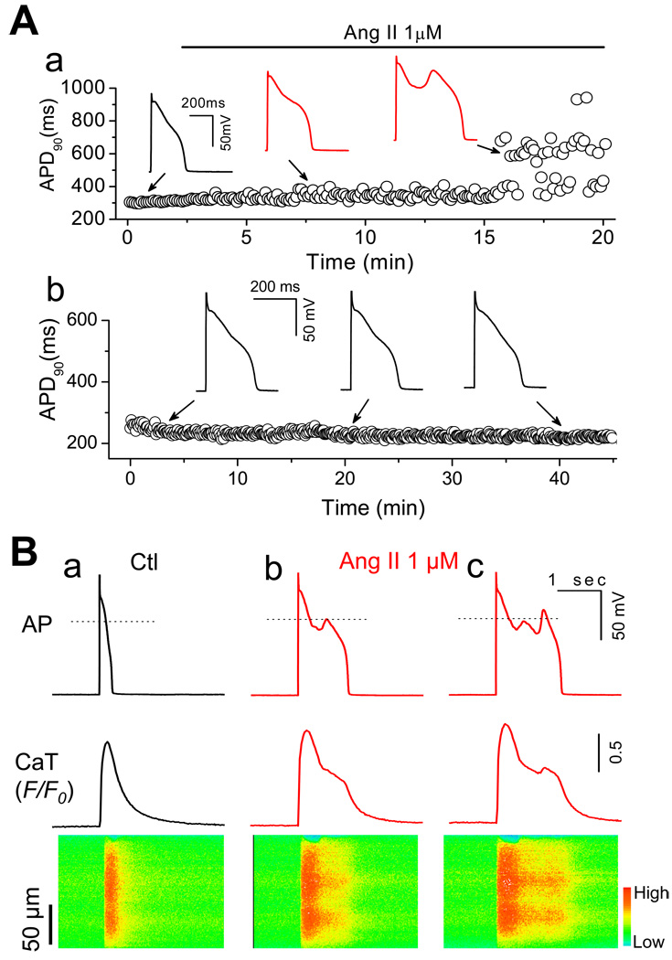
Fig. 2.
Early afterdepolarizations (EADs) and intracellular calcium (Cai) alteration induced by Ang II. (A-a). APs recorded under perforated whole-cell configuration before and during Ang II perfusion. Values of consecutive APD90 are plotted over time. EADs were induced at 15.8 ± 1.6 min after exposure to Ang II. (A-b). A representative AP recording from a cell perfused with control perfusate for > 40 min. No EADs were observed. (B). Cai Transients recorded under control and during Ang II-Induced EADs. a. AP, whole-cell Cai transient, and a line-scan image along the long axis of the myocyte before Ang II treatment. b & c. Same following exposure to 1µM Ang II for ~20 min. EADs result in persistent elevation (b) and additional release in Cai (c).

