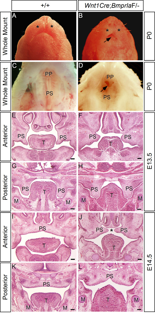
Figure 2.
Wnt1Cre;BmprIaF/− mice exhibit a unique anterior clefting of the secondary palate. (A-D) Gross examination of wild type (A, C) and Wnt1Cre;BmprIaF/− (B, D) mice at postnatal day 0 (P0) reveals craniofacial defects, including extremely shortened mandible and hypoplastic maxillary prominence (asterisks) (B), and an unusual type of anterior clefting of the secondary palate (arrow) (D). (E-H) coronal sections of E13.5 wild type control (E, G) and Wnt1Cre;BmprIaF/− (F, H) embryos reveal shortened and horizontally positioned palatal shelves in the anterior region of mutant (F). The posterior palatal shelves appear morphologically comparable in wild type control and mutant ( G, H). (I-L) At E14.5 when the palatal shelves meet and fuse at the midline at the anterior (I) and posterior (K) domains in wild type control, the mutant palatal shelves appear too short to meet in the anterior region (J), but do meet and fuse in the posterior region (L). M, Meckel’s cartilage; T, tongue; PP, primary palate; PS, palatal shelf. Asterisks in (A) and (B) mark maxillary prominence. Asterisk in (J) marks palate clefting. Arrow in (B) points to exposed tongue, and in (D) points to the anterior clefting of the secondary palate. Scale bars represent 100 µm.

