Figure 1.
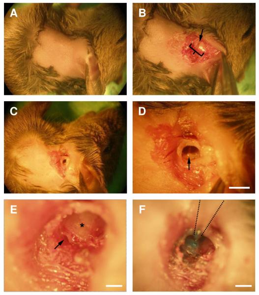
Demonstration of the surgical approach in a euthanized animal. The animals lies on its right side, facing to the right, with its ventral side to the top of the field of view.
A. Hair from a small patch of postauricular skin was removed.
B. Incision was made in the postauricular area. Soft tissue was retracted to reveal the ear canal (bracket) and an incision was made in the clear area in the middle of the ear canal (arrow).
C. A small cut was made in the ear canal.
D. View of the tympanic membrane and the malleus (arrow). Scale bar = 1 mm.
E. Exposed view of round window (arrow) after removing part of the bulla. Asterisk – band shows the division point between the two turns of the cochlea. A hole is made in the cochlear wall just ventral to this. Scale bar = 0.1 mm.
F. A micropipette containing dye (Fast Green) was inserted into a hole drilled in the cochlea wall and the dye was pressure-injected. Scale bar = 0.2 mm.

