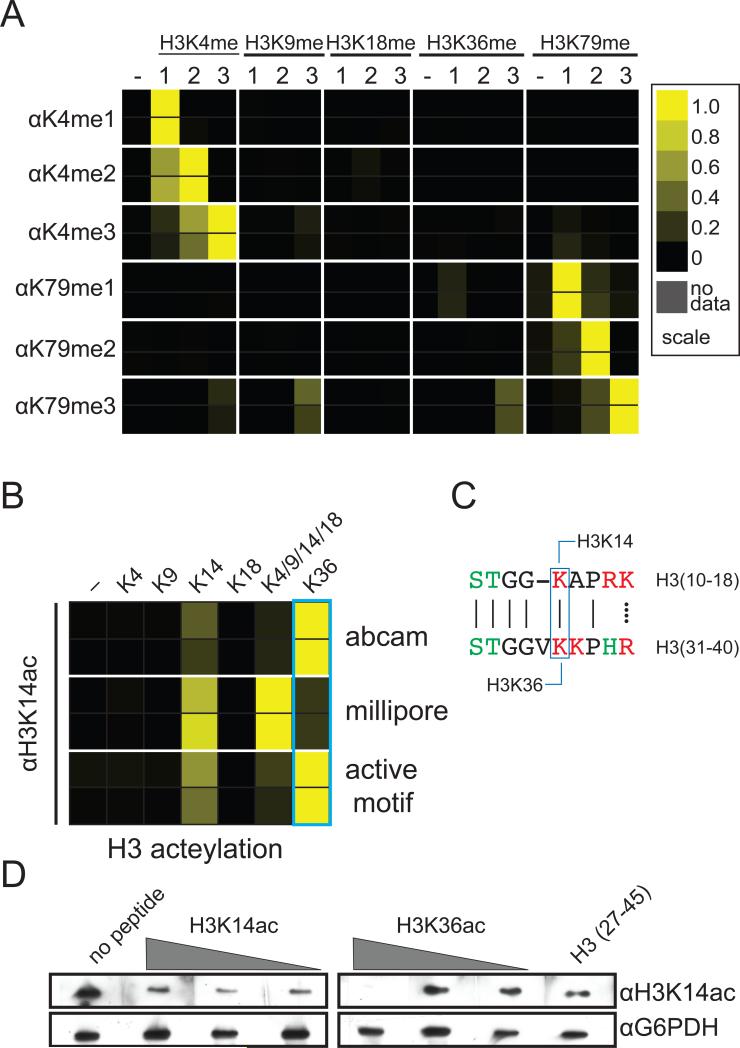
Figure 2.
Antibody binding to histone peptide microarrays. Results of two independent arrays consisting of 24 independent spots for each peptide are depicted as heatmaps of the normalized mean intensity and plotted on a scale from 0 to 1 with 1 (yellow) being the most significant (see Methods). (A) Interactions of H3K4- and H3K79-specific antibodies with methylated peptides derived from the N-terminus of histone H3 (antibodies used are given in Table S1 and further information in Figure S1 and S3). (B) Recognition of histone H3 acetyllysine peptides by H3K14ac antibodies. (C) Alignment of sequence surrounding H3K14 and H3K16. (D) Western blot of yeast whole cell extract probed with H3K14ac antibody preincubated with various concentrations of histone H3 peptides.

