Fig. 3.
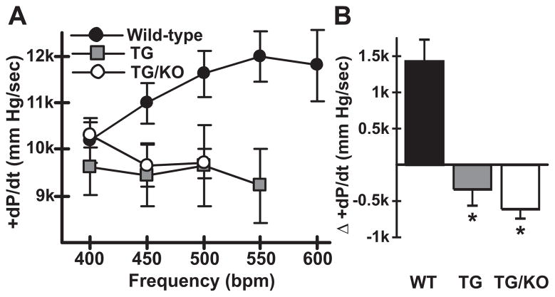
TM180 (TG) and TM180/AE3 (TG/KO) mutants exhibit a negative force-frequency response. Hearts of anesthetized surgically-instrumented 3-month-old mice were subjected to atrial pacing beginning at 400 beats per minute (bpm) and contractile parameters were measured. n = 5 WT, 4 TG, and 5 TG/KO mice, with 2 males and either 2 or 3 females of each genotype. WT mice could be paced to 550 and 600 bpm but some TG and TG/KO mice could not. If fewer than 3 mice could achieve a given frequency, such as TG/KO at 550 bpm, the data were not plotted. (A) A positive FFR with respect to maximum +dP/dt (mm Hg/sec) was observed in WT mice but not in TG and TG/KO mice. (B) Difference in +dP/dt at 400 bpm and 500 bpm revealed a negative FFR in TG and TG/KO mice. *p < 0.02 vs WT.

