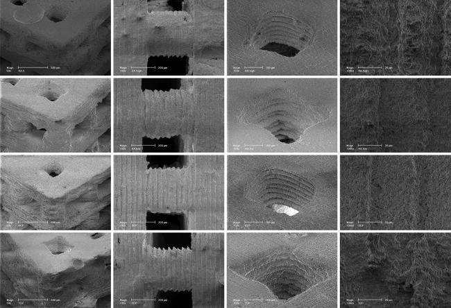
Fig. 3.
SEM micrographs of the four scaffold materials. Rows top to bottom HA h, HA l, BCP, and TCP. First column Perspective view of scaffolds at 50× magnification (bar 50 µm). Second column Scaffold structures at 100× magnification (bar 200 µm). Note the regular surface texture on the scaffolds. Third column Scaffold pores at 150× magnification (bar 200 µm). Fourth column Scaffold surfaces at 1000× magnification (bar 20 µm)

