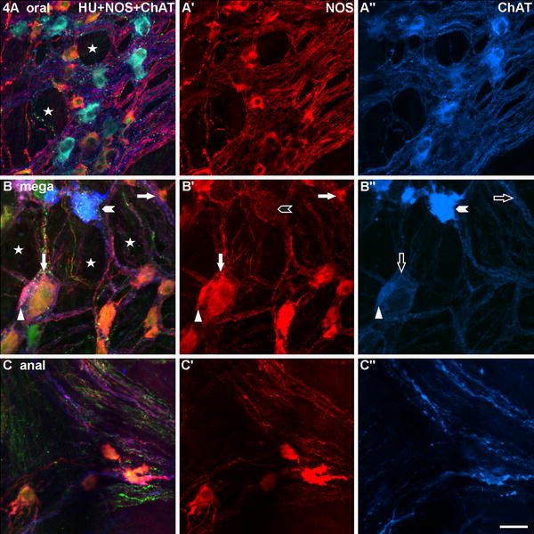
Fig. 4.

Three myenteric ganglia gained from wholemounts of the megacolonic, seropositive, female patient aged 69 years, stained for human neuronal protein Hu C/D (HU), neuronal nitric oxide synthase (NOS) and common choline acetyltransferase (ChAT). a is from the oral, b from the dilated (mega), c from the anal part of the resected specimen. The ganglion in a appears, though some honeycomb-like structures (asterisks), still normal, the ganglion in b is strikingly bulked (asterisks). In a, there is a quite balanced quantitative relation between NOS- and ChAT-neurons, in b and c, the ganglia contain predominantly NOS-neurons (exemplarily: arrows). Mainly in b, the NOS-neurons differ considerably in size (compare the two arrowed neurons). Two marked ChAT-neurons in b″ differ markedly in their staining intensities (compare the arrowheaded with the triangled neuron, the latter is additionally co-reactive for NOS and HU: triangles). Bar 50 μm
