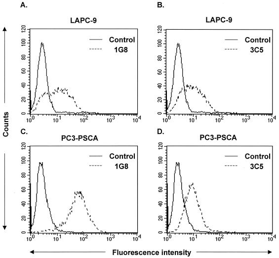
Figure 1.
Detection of cell-surface PSCA expression by anti-PSCA mAbs using flow cytometry. (A and B) LAPC-9 cells were stained with either 1G8 (A) or 3C5 (B) mAbs (dotted line) and the corresponding isotype control antibodies (solid line) IgG1 (A) or IgG2a (B). (C and D) PC3-PSCA cells were stained with either 1G8 (C) or 3C5 (D) mAbs (dotted line). As a control, PC3 cells (solid line) were stained with 1G8 (C) or 3C5 (D).

