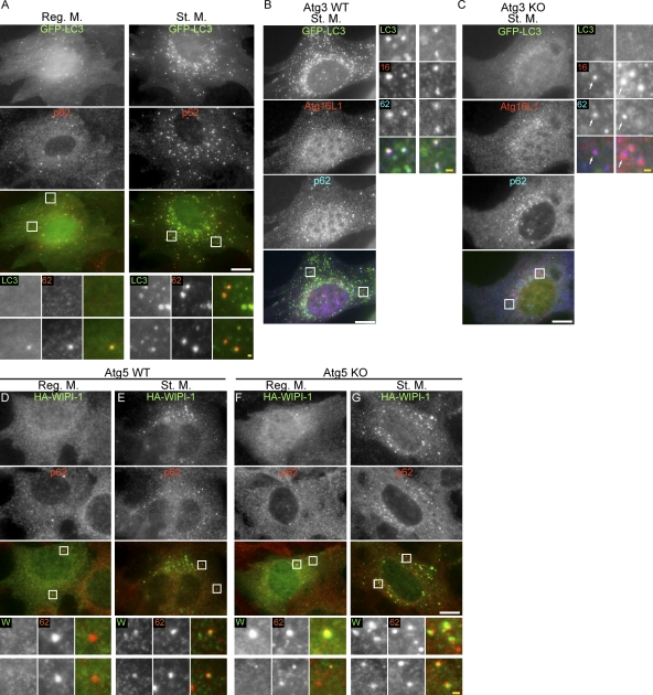
Figure 1.
Localization of p62 to the early autophagic structures is independent of LC3. (A) MEFs stably expressing GFP-LC3 were cultured in regular medium or starvation medium for 1 h. Cells were stained with anti-p62 antibodies and analyzed by immunofluorescence microscopy. (B and C) Localization of p62 to the autophagic structures is independent of LC3 lipidation. Wild-type (B) and Atg3 KO MEFs (C) stably expressing GFP-LC3 were cultured in starvation medium for 2 h. Cells were stained with anti-Atg16L1 and anti-p62 antibodies. Structures positive for Atg16L1 and p62 but not for GFP-LC3 are indicated by arrows. (D–G) Localization of p62 to the autophagic structures is independent of Atg5. Wild-type (D and E) and Atg5 KO MEFs (F and G) stably expressing HA–WIPI-1 were cultured in regular (D and F) or starvation medium (E and G) for 2 h. Cells were stained with anti-HA and anti-p62 antibodies. Signal color is indicated by color of typeface. Reg. M., regular medium; St. M., starvation medium; WT, wild type. Bars: (white) 10 µm; (yellow) 1 µm.

