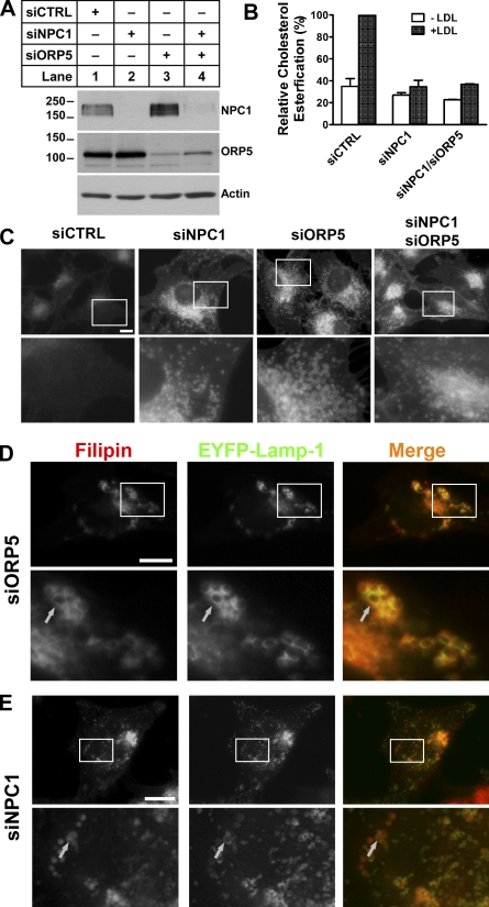
Figure 4.
Cholesterol accumulates on the limiting membrane of LE/LY in ORP5 knockdown cells. (A) HeLa cells grown in medium A were transfected with siNPC1, siORP5, siNPC1, and siORP5, or a universal control siCTRL for 48 h, followed by transfection with pcDNA4-ORP5 for 24 h. Efficiency of knockdown was analyzed by immunoblotting using polyclonal anti-NPC1, anti-ORP5 antibodies. The molecular mass of the proteins is indicated in kilodaltons next to the gel blot. (B) HeLa cells grown in medium A were transfected with the indicated siRNAs for 48 h, followed by incubation in medium D for 18 h. Cells were then chased with 50 µg/ml LDL and [14C]-palmitate in medium D for 6 h. A cholesterol esterification assay was performed as described in Fig. 3 D, and relative cholesterol esterification was quantified as described in Fig. 3 E. The results are expressed as means + SD (error bars). (C) HeLa cells grown in medium A were transfected with indicated siRNAs for 54 h. Cells then received medium D supplemented with 50 µg/ml LDL for 18 h followed by processing for filipin staining. Fluorescent images are representative of four independent experiments with similar results. Bars, 10 µm. (D and E) HeLa cells grown in medium A were transfected with siORP5 (D) or siNPC1 (E) for 48 h, followed by transfection with pEYFP-Lamp1 for 6 h. Cells then received medium D supplemented with 50 µg/ml LDL for 18 h followed by processing for filipin staining and fluorescence microscopy. Enlarged views of the boxed regions are shown in the bottom panels. Arrows indicate the overlapping of accumulated cholesterol with EYFP-Lamp-1 in ORP5 knockdown cells but not in NPC1 knockdown cells. Bars, 10 µm.

