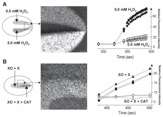
Fig. 4.
Multiple injury experiments conducted in DCFH-loaded cardiac myocyte cultures. (A) This experiment was conducted using a double injury chamber. The two injury zones were perfused with the Tyrode solution containing 0.5 mM and 5 mM H2O2. Traces on the graph show rapid increase in fluorescence levels associated with the transition from DCFH to DCF. The traces were acquired from three different regions of interest positioned within each zone. The image illustrates large differences in fluorescence levels between the two areas and the control network. (B) This experiment was conducted using a nested injury chamber. The large injury zone was perfused with 0.2 U/mL xanthine oxidase and 1 mM xanthine, whereas the solution flowing via the small injury zone also included an additional 400 U/mL of catalase. The difference between the two areas illustrates the inability of superoxide anions to oxidize intracellular DCFH.

