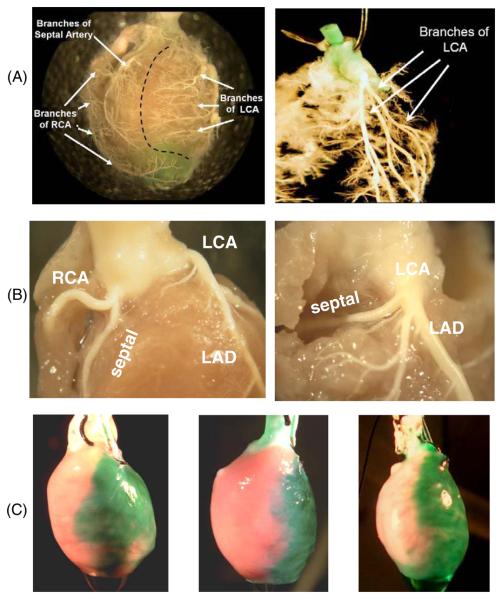
Figure 1.
Coronary vessel structure in the rat. (A) Right and left coronary artery beds. Polymer casts show the main two branches of the coronary circulation. RCA: right coronary artery. LCA: left coronary artery. (B) Branches of the RCA and LCA near the ostium. The LAD is the main branch off the LCA. A case of the septal artery branching from the RCA is shown on the left and a case of the septal artery branching from the LCA is shown on the right. (Panel C) injection of green dye into the microcannula reveals the tissue bed fed by the LAD. Three microcannulated hearts are shown. The anatomies of individual coronary beds are slightly different between hearts and so are the areas of affected epicardial tissue.

