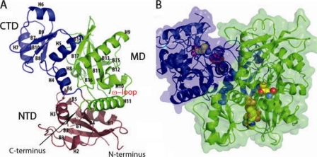
FIG. 2.
Overall structure of M. tuberculosis Ddl and domain definition. (A) Ribbon diagram of the Ddl monomer, with helices (H) as well as β-sheet regions (B) defined. Domains are colored blue (CTD), green (MD), and purple (NTD). (B) Transparent space-filling representation of the Ddl dimer, with a ribbon diagram shown beneath the surface. ADP and d-Ala:d-Ala dipeptide are shown as spheres colored by atom type, which were docked in manually, using the ligand-bound form of E. coli Ddlb (PDB accession code 1IOV) as a guide (14). Monomers are denoted by different colors (green and purple).

