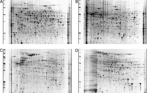
FIG. 2.
2D gels of cellular fractions. All subcellular fractions were subjected to 2-dimensional gel electrophoresis using strips of pI 5 to 8. (A) The cytoplasmic fraction tends to have the highest quantity of proteins of the four fractions. (B and C) The microsomal (B) and cell wall/plasma membrane (C) fractions may have some overlap due to incomplete separation. (D) Secreted proteins tend to be the fewest in number. All 4 fractions have individual patterns and proteins that are unique to that fraction alone as well as overlapping proteins that are consistent across fractions. All gels were stained with SYPRO ruby for visualization.

