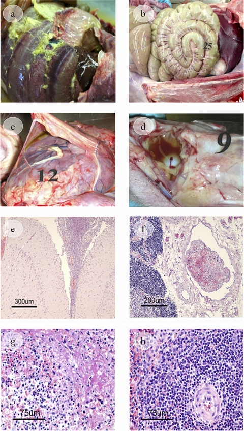
FIG. 4.
Gross and histopathological findings. Images in the peritoneal cavity show severe fibrinous peritonitis (nonvaccinated control piglet) (a) and mild fibrinous peritonitis (NPAPTM-vaccinated piglet) (b). (c) Image of the pericardial cavity and heart showing a fibrinous pericarditis (rTbpA piglet). (d) Image of the hock joint showing fibrinous arthritis (inflammatory fluid and fibrin strands, rTbpB piglet). (e) Image of the meninges showing fibrinous-suppurative meningitis. Note a thickening of the meninges covering the brain due to the presence of fibrin and inflammatory cells in the subarachnoid space (H&E) (rTbpA piglet). (f) Image of a blood vessel showing vascular thrombosis (H&E) (rTbpB piglet). (g) An image of spleen tissue shows lympholysis of the follicular center cells, with the production of nuclear debris. Transudation of plasma proteins is also seen (H&E) (nonvaccinated control piglet). (h) No necrotic lymphocytes of the white pulp are seen in spleen tissue (H&E) (NPAPTCp piglet).

