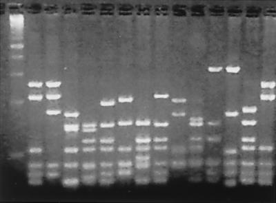
Figure 3.
DNA fingerprint analysis of the scFv genes of individual antibodies from the second round of selection. scFv DNA was amplified by PCR directly from colonies and digested with the frequently cutting restriction enzyme BstN1. A diverse banding pattern was observed, with each unique pattern representing a unique antibody sequence. First lane is a 100-bp DNA marker.

