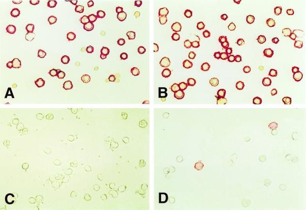
Figure 4.
Staining of adult buffy coat WBCs and total fetal liver by phage antibodies. Adult buffy coat WBCs or fetal liver cells were stained with biotinylated phage antibodies and the cells applied to microscope slides, fixed, and stained with streptavidin-alkaline phosphatase and Fast Red. The phage antibody FSA7 stains fetal erythroid cells (A) but not WBCs (C), whereas the phage antibody FSH3 stains both fetal liver (B) and a subset of WBCs (D).

