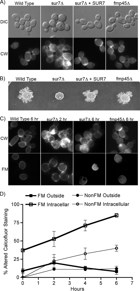
Fig. 2.
Analysis of the cell wall and morphogenesis phenotypes of the sur7Δ and fmp45Δ mutants. (A) The morphology of live cells of the indicated genotypes is shown in the upper images. The lower images show a different set of cells, viewed by fluorescence microscopy, that were permeabilized and stained with calcofluor white to detect cell wall structures. (B) Invasive-growth assay of the indicated C. albicans strains. Equal numbers of cells were spotted onto an agar plate containing 4% serum and then incubated at 37°C for 7 days. (C) Comparison of cell wall structures in older and newer cells. Cells of the indicated genotype were cross-linked to fluorescein-maleimide and then grown for 2 or 6 h at 30°C in YPD. The cells were then permeabilized and stained with calcofluor white. Fluorescence microscope images of cells stained with calcofluor white are shown above; the older cells that were cross-linked with fluorescein are shown below. (D) Quantitation of calcofluor white-stained cells after labeling with FM at time zero versus new cells that were not labeled (NonFM). Outside, cells that showed altered calcofluor white staining of the outer cell wall; Intracellular, altered calcofluor white staining within the cell due to ectopic growth of the cell wall. The error bars indicate SD.

