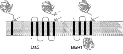
FIG. 4.
Topology of SPase substrates. (Left) General representation of a signal peptide protein showing a transmembrane region and an extracellular domain that is released via the indicated SPase cleavage site (arrow). (Center) Proposed topology of LtaS showing five TM domains and two proposed cleavage sites, one (solid arrow) predicted by SignalP 3.0 (9) and a second (hashed arrow) predicted by proteomic data (36, 66). (Right) Proposed topology of BlaR1 (20) showing four TM domains, an intracellular zinc binding domain, and the extracellular domain released via the indicated cleavage site.

