FIG. 5.
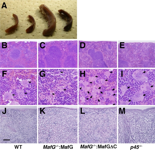
Accumulation of megakaryocytes and fibrosis observed in spleens of MafG−/−::G1HRD-MafGΔC mice. (A) Macroscopic observation of spleens. Spleens of (from the left) wild-type, MafG−/−::G1HRD-MafG line 8, MafG−/−::G1HRD-MafGΔC line 54, and MafG−/−::G1HRD-MafGΔC line 36 mice are shown. (B to I) Hematoxylin-and-eosin (HE) staining of spleens of wild-type (B and F), MafG−/−::G1HRD-MafG line 8 (C and G), MafG−/−::G1HRD-MafGΔC line 36 (D and H), and p45-null (E and I) mice. Arrows indicate megakaryocytes. (J to M) Silver impregnation of spleens of wild-type (J), MafG−/−::G1HRD-MafG line 8 (K), MafG−/−::G1HRD-MafGΔC line 36 (L), or p45-null (M) mice. Scale bar, 500 μm (B to E), 50 μm (F to I), or 200 μm (J to M).

