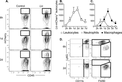
FIG. 3.
Progression of L. infantum chagasi parasites through different inflammatory cell types during the first 7 days after introduction into host tissues. BALB/c mice were inoculated intradermally in the ear with 106 transgenic metacyclic L. infantum chagasi promastigotes expressing the fluorescent protein mCherry and with saline as a control. Dermal inflammatory cells harboring intracellular parasites were characterized by flow cytometry with antibodies to surface markers. (A) Representative contour plot from three time points after parasite inoculation are shown. There is a progression of CD45+ cells harboring fluorescent L. infantum chagasi in inflammatory cells from mouse ears inoculated with parasites but not saline controls. The total numbers of L. infantum chagasi-containing leukocytes that stain for CD45+ are shown in panel B, and the CD45+ cells that costain for markers of neutrophils (Ly6G) or macrophages (CD11b+ CD11c− Ly6G−) are indicated in panel C. (D) Cells were gated on dermal inflammatory CD45+ leukocytes that costain for mCherry. Plots show subpopulations of these L. infantum chagasi-harboring cells that express surface markers for neutrophils and/or macrophages, as in panel C.

