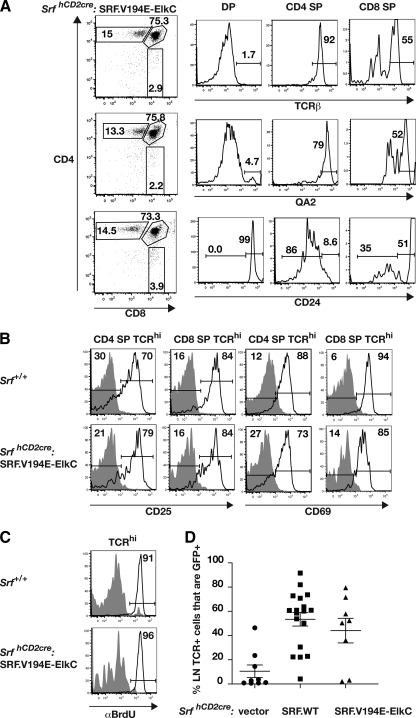
FIG. 4.
The SRF-Elk fusion protein generates mature thymocytes that can be activated through the T-cell receptor. SrfhCD2cre:SRF.V194E-ElkC thymocytes were analyzed from reconstituted Rag2−/− animals by gating on GFP+ cells. (A) TCR-β, QA2, and CD25 expression during thymocyte development. SrfhCD2cre:SRF.V194E-ElkC thymocytes were gated on DP, CD4 SP, and CD8 SP cells and analyzed for expression of the indicated markers. (B) Upregulation of CD25 and CD69 following TCR cross-linking. Wild-type (top panels) or SrfhCD2cre:SRF.V194E-ElkC thymocytes were analyzed before (gray shading) or 48 h after (black lines) stimulation by anti-CD3/anti-CD28. (C) TCR cross-linking induces proliferation of SrfhCD2cre:SRF.V194E-ElkC thymocytes. Proliferation was assessed by incorporation of BrdU during a 20-h pulse either without stimulation (gray shading) or 48-h after stimulation by anti-CD3 and anti-CD28. (Top) Wild-type thymocytes; (bottom) SrfhCD2cre:SRF.V194E-ElkC thymocytes. (D) Proportions of GFP+ (i.e., retrovirally transduced) lymph node TCR+ cells in animals reconstituted with SrfhCD2cre cells transduced by vector, wild-type SRF, or SRF.V194E-ElkC.

