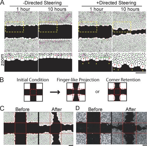
FIG. 2.
Steering model predicting collective endothelial migration behaviors. (A) Computational cell monolayers respond to a large open space over 10 simulated hours in the absence (left) and presence (right) of growth factor. Enlargements of the regions outlined by yellow dashes are shown below. The colors of the nuclei indicate the dominant steering type: green, adhesive; red, directed; magenta, repulsive; and black, lateral drag. The arrows illustrate the direction and magnitude of steering terms. (B) Schematic representation of two possible migration outcomes for monolayers starting with triangular projections: finger extension versus corner retention. (C) Images of simulated monolayers responding to a cell-free area with pioneer cells at artificially generated triangular corner positions. The snapshots represent cells immediately after the introduction of cell-free space and after a simulated 12-h period, showing corner retention. (D) HUVEC subjected to a cross scratch that generated triangular projections before and after a 12-h incubation period, showing corner retention. The cells were assayed as in Fig. 1A. Bars, 150 μm.

