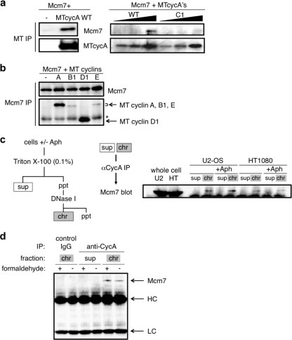
FIG. 3.
Interaction of cyclin A and Mcm7 in human cells. (a) 293 cells were cotransfected with 5 μg of Mcm7- and MT cyclin A (5 μg in the left panel and 1, 2, and 4 μg in the right panel for the wild type; 1, 2, and 4 μg in the right panel for C1)-expressing vectors, and the cell extracts were subjected to anti-myc tag immunoprecipitation (IP), followed by anti-Mcm7 and anti-MT Western blot analysis. (b) 293 cells were cotransfected with 5 μg each of Mcm7- and myc-tagged-cyclin (cyclin A, cyclin B1, cyclin D1, and cyclin E)-expressing vectors, and the cell extracts were subjected to anti-Mcm7 immunoprecipitation, followed by anti-Mcm7 and anti-myc tag Western blot analysis. The asterisk indicates the IgG heavy chain. (c) Cell extracts from asynchronous or aphidicolin (Aph)-blocked U2-OS and HT1080 cells were fractionated into soluble (sup) and chromatin-bound (chr) fractions and subjected to anti-cyclin A (αCycA) immunoprecipitation, followed by anti-Mcm7 Western blot analysis. (d) Cell extracts from asynchronous U2-OS cells treated with (+) or without (-) 0.2% formaldehyde were fractionated into soluble and chromatin-bound fractions as for panel c and subjected to anti-cyclin A (rabbit polyclonal) immunoprecipitation, followed by anti-Mcm7 (rabbit polyclonal) Western blot analysis. The chromatin-bound fractions were also immunoprecipitated with control rabbit IgG. Arrows indicate Mcm7, the IgG heavy chain (HC), and the IgG light chain (LC). ppt, precipitates.

