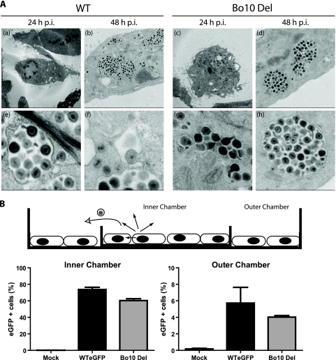
FIG. 5.
The Bo10 deletion phenotype is not associated with a virion release deficit. (A) Electron microscopy of virus-infected cells. MDBK cells were infected (24 or 48 h; 1 PFU/cell) with WT or Bo10 Del virus as indicated. The pictures shown are representative of at least 10 sections per sample. For each time point, two different pictures at different magnifications are provided. (B) Fluid-phase virus spread. A total of 2 × 10⁵ infected MDBK cells (MOI of 0.5) or 2 × 10⁵ uninfected MDBK cells were seeded either on a 6-cm-diameter dish or in a 3.5-cm-diameter dish placed inside the first one. The cells were therefore separated by a physical barrier but connected by medium so that virus could not spread directly from the infected to the uninfected population but only via their common supernatant. After 48 h, the cells were harvested and analyzed for eGFP expression (eGFP + cells, eGFP-expressing cells). The data presented are the averages plus SEMs (error bars) for triplicate measurements and were analyzed by t test. The values for the WT eGFP and Bo10 Del virus strains were not significantly different.

