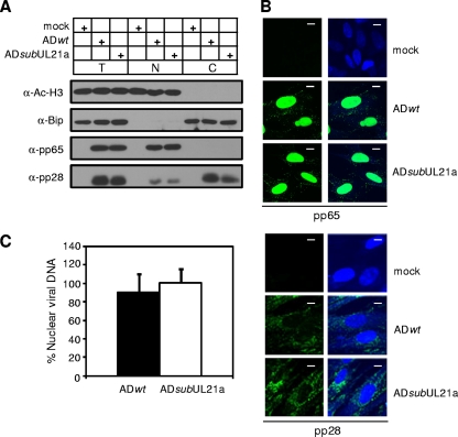
FIG. 2.
UL21a deletion virus is competent for viral entry. (A and B) Translocation of tegument proteins pp65 and pp28 delivered into infected cells. MRC-5 cells were infected with ADwt and ADsubUL21a at an input genome number equivalent to 1 or 10 TCID50 units of wild-type virus/cell, collected at 2 hpi, and analyzed by immunoblotting (A) or immunofluorescence (B), respectively. For immunoblotting, total lysate (T), nuclear (N), and cytoplasmic (C) fractions were prepared and analyzed as indicated. Acetyl-histone H3 and Bip were used as nuclear and cytoplasmic fractionation controls, respectively. For immunofluorescence, the staining patterns of nuclei and viral antigens are shown as blue and green, respectively. Scale bar, 10 μm. (C) Nuclear translocation of virion DNA. Intracellular DNA was isolated from both total and nuclear fractions at 2 hpi. The quantity of viral genomes in each fraction was determined by qPCR using primers specific for the viral UL54 gene, and viral genome copies were normalized to cellular genomes using β-actin primers. The percentage of viral DNA translocated into the nucleus was determined by dividing viral genome equivalents in the nuclear fraction by those in the total cell lysate. Shown are representative results from at least two independent experiments. Error bars indicate standard deviations.

