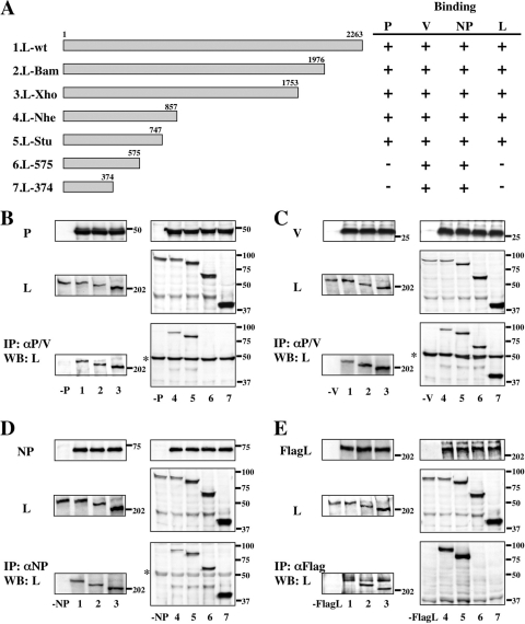
FIG. 1.
Analysis of interactions between the C-terminally truncated L and P, V, NP, or L proteins by immunoprecipitation (IP) and Western blot (WB) assay. (A) Schematic diagram of the C-terminally truncated L proteins. Binding to P, V, NP, or L protein is summarized at right. (B to E) The proteins from transfected BSR T7/5 cell lysates were analyzed by Western blotting for anti-P/V MAb (B and C), anti-NP MAb (D), or anti-Flag polyclonal antibody (E) (top panels) and anti-L MAb (middle panels). Immunoprecipitates with anti-P/V MAb (B and C), anti-NP MAb (D), or anti-Flag antibody (E) were probed by anti-L MAb in a Western blot assay (bottom panels). Numbers on the bottom correspond to the L proteins depicted in panel A. Sizes of molecular mass markers (in kDa) are shown. The asterisk at the left indicates the immunoglobulin heavy chain.

