FIG. 2.
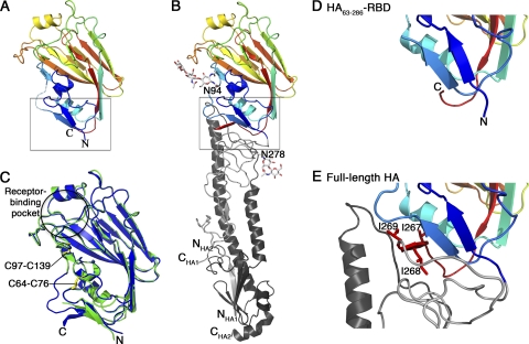
Structural comparison of the HA receptor-binding domain in the HA63-286-RBD (A) and the A/H1N1/2009 HA ectodomain (B). In these two panels, the domain is colored blue to red, N to C terminus, to facilitate the chain trace. Glycosylation at Asn94 and Asn278 in the HA ectodomain is shown as a ball-and-stick model. (C) Overlay of HA63-286-RBD (green) and equivalent residues in HA ectodomain (blue). The locations of the receptor-binding pocket and two disulfide bonds are highlighted. (D and E) Detail of boxed regions in panels A and B, respectively, corresponding to the interface region between the receptor-binding domain and the helical stalk region that undergoes pH-dependent conformational changes. Note that the isoleucine-rich third β-strand of the interfacial β-sheet in panel E is disordered in panel D. The coordinates for HA63-286-RBD have been deposited at the Protein Data Bank as PDB entry 3MLH. The coordinates for the A/H1N1/2009 HA ectodomain are from PDB entry 3LZG. All residues are labeled using H3 numbering.

