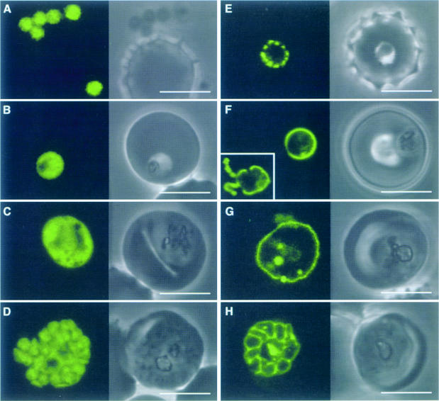
Fig. 3. Plasmodium falciparum expressing ACPtransit–GFP (A–D) and ACPsignal–GFP (E–H). ACPtransit–GFP expression results in cytoplasmic GFP in merozoites (A) through to schizonts (D). GFP is apparently excluded from the food vacuole (B and C) and other organelles (C). ACPsignal–GFP expression results in GFP export to the parasitophorous vacuolar space (in which the parasite resides within the erythrocyte, compare with Figure 2D). Newly invaded ring parasites (E) show punctate fluorescence in the parasitophorous vacuole, which may indicate some early formative stage of this specialized structure. Subsequent stages show continuous fluorescence surrounding the parasite (F and G), which ultimately delineates the products of schizogony (H). GFP is also seen in the tubulovesicular network [(F), inset, fluorescence only]. Some internalized GFP probably represents protein that re-enters the parasite in food vacuoles. Scale bars, 5 µm.

An official website of the United States government
Here's how you know
Official websites use .gov
A
.gov website belongs to an official
government organization in the United States.
Secure .gov websites use HTTPS
A lock (
) or https:// means you've safely
connected to the .gov website. Share sensitive
information only on official, secure websites.
