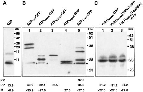
Fig. 4. Western blot analyses of apicoplast-targeted proteins using ACP antiserum to detect endogenous ACP (A) or GFP antiserum to detect ACP–GFP-based fusion proteins (B) and FabH–GFP-based fusion proteins (C). Bands marked with ‘*’ indicate GFP degradation products. Predicted sizes of mature (‘M’) and pre-processed (‘PP’) forms of each protein as discussed in the text are shown below each lane in kDa. Sizes marked with ‘>’ indicate minimal estimates due to uncertainty of the precise processing point.

An official website of the United States government
Here's how you know
Official websites use .gov
A
.gov website belongs to an official
government organization in the United States.
Secure .gov websites use HTTPS
A lock (
) or https:// means you've safely
connected to the .gov website. Share sensitive
information only on official, secure websites.
