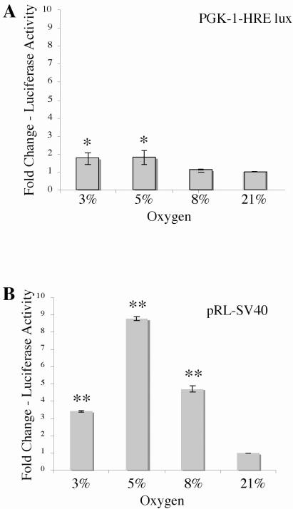
Figure 1. Induction of PGK-1-HRE and pRL-SV40 luciferase reporters by hypoxia.
In the conventional luciferase assay, two or more plates are plated with equal numbers of cells and individually transfected with an experimental and a constitutive luciferase reporter construct. During analysis, differences in initial transfection efficiency are accounted for by normalizing the luminescence obtained for the experimental reporter to the luminescence obtained for the constitutive reporter. Normalized reporter luminescence under experimental conditions is then compared with that of the control condition to obtain results as fold change over control. Rcho-1 trophoblast cells were plated at equal cell number of 1.0 × 10⁵ cells and transfected with 1 μg PGK-1-HRE and 0.2μg pRL-SV40 and incubated at the O2 concentration indicated for 18hrs and analyzed for (A) PGK-1-HRE reporter activation and (B) pRL-SV40 constitutive reporter activation. Results are the average of three independent experiments. Luciferase activity at each concentration was analyzed and is shown relative to activity at 21% oxygen. Error bars represent standard deviation. Significance was denoted as *p≤0.01 or **p≤0.001 and was determined using a one way Anova with Tukeys post hoc test.

