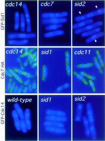
Fig. 4. Dependencies for localization of Sid1p, Cdc14p and Cdc7p. The localizations of GFP–Sid1p, Cdc7-HA and GFP–Cdc14p were examined in wild-type and different sid mutant cells after incubation at 36°C for 2 h. All samples were fixed and stained for DNA (blue), GFP–Sid1p and GFP–Cdc14p signal, and Cdc7-HA staining is shown in green. Cdc7-HA cells have a bright green haze as a result of background staining from the secondary antibody.

An official website of the United States government
Here's how you know
Official websites use .gov
A
.gov website belongs to an official
government organization in the United States.
Secure .gov websites use HTTPS
A lock (
) or https:// means you've safely
connected to the .gov website. Share sensitive
information only on official, secure websites.
