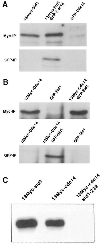
Fig. 5. Sid1p and Cdc14p associate in vivo. (A) Lysates were prepared from cells expressing 13Myc-Sid1p (YDM617), GFP–Cdc14p (YDM627) or both (YDM629). (B) Lysates were prepared from cells expressing 13Myc-Cdc14p (YDM617), GFP–Sid1p (YDM558) or both (YDM626). Each lysate was split in two, and immunoprecipitations were performed using anti-Myc or anti-GFP antibodies. Immune complexes were analyzed by Western blotting using anti-Myc antibodies. (C) Wild-type cells expressing 13Myc-Sid1p (YDM587) or 13Myc-Cdc14p (YDM617), or sid1-239 (YDM637) cells expressing 13Myc-Cdc14p were started at 25°C, shifted to 36°C for 3 h, and kinase assays were performed on 13Myc-Cdc14 or 13Myc-Sid1 immune complexes from these cells.

An official website of the United States government
Here's how you know
Official websites use .gov
A
.gov website belongs to an official
government organization in the United States.
Secure .gov websites use HTTPS
A lock (
) or https:// means you've safely
connected to the .gov website. Share sensitive
information only on official, secure websites.
