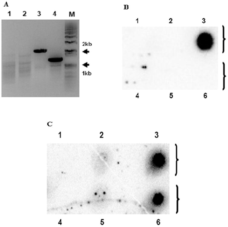
Fig. 4.

The kanR marker was replaced with the zeoR marker during serial passage of ΔIR3 EHV-1. PCR and dot blot hybridization analyses were performed as described in Materials and Methods. (A) PCR amplification of flanking regions of markers inserted into ΔIR3 EHV-1 genome. ΔIR3 EHV-1 infected RK13 DNA was used as template. KanR left flanking region amplification primer set (lane 1). KanR right flanking region amplification primer set (lane 2). ZeoR left flanking region amplification primer set (lane 3). ZeoR right flanking region amplification primer set (lane 4). “M” is DNA size marker. (B and C) Dot blot hybridization analyses with probes specific for the kanR and zeoR markers, respectively. Lanes 1 to 6 represent mock DNA, pRacL11 EHV-1 DNA, pΔIR3 EHV-1 DNA, RK13 DNA, RacL11 EHV-1 infected RK13 DNA, and ΔIR3 EHV-1 infected RK13 DNA, respectively.
