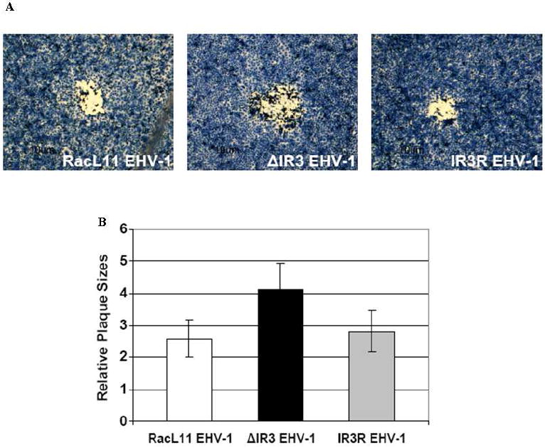
Fig. 5.

Comparison of plaque size of RacL11 EHV-1, ΔIR3 EHV-1 and IR3R EHV-1 in RK13 cells. (A) Morphology of plaques in RK13 cells infected with RacL11 EHV-1, ΔIR3 EHV-1 and IR3R EHV-1. (B) Comparison of plaque size in RK13 cells infected with RacL11 EHV-1, ΔIR3 EHV-1 and IR3R EHV-1. Relative plaque sizes were measured by using the ImageJ software program; the two-tailed Student’s t test was used for data analysis. Error bars indicate standard deviation.
