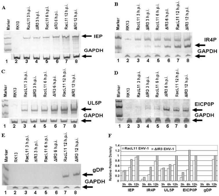
Fig. 7.

Expression levels of EHV-1 IE, early, and late proteins. RK13 cells infected with ΔIR3 EHV-1 or RacL11 EHV-1 at a moi of 10 were harvested at 3, 6, and 12 hpi, and protein expression was examined by western blot analysis as described in Materials and Methods. Detection of the (A) immediate early protein, (B) early IR4 protein, (C) early UL5 protein, (D) early EICP0 protein, and (E) late glycoprotein. (F) Relative protein density of EHV-1 representative genes in RacL11 and ΔIR3 EHV-1 infected RK13 cells. Protein density was compared by using the ImageJ software program (http://rebweb.nih.gov/ij/). White and gray bars indicate proteins of RK13 cells infected with RacL11 EHV-1 and ΔIR3 EHV-1, respectively. Relative protein density was the protein density of each protein relative to the amount of that protein in RK13 cells infected with ΔIR3 EHV-1 at 12hpi.
