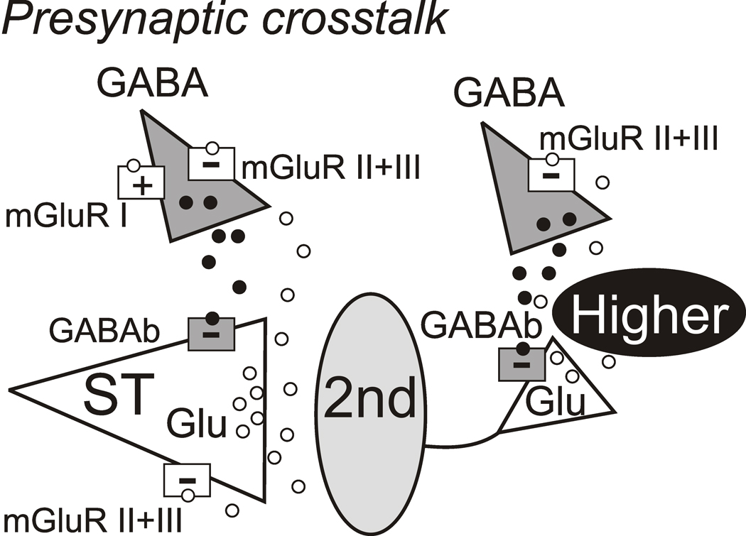
Figure 6.
Summary of functioning presynaptic distributions of mGluR subtypes and GABAB on synaptic terminals in second-order and higher order NTS neurons. Glutamate (Glu) mechanisms are white with triangles representing synapses and GABA mechanisms are gray. Released glutamate (unfilled circles) and GABA vesicles (filled circles) are within diffusion distance of glutamate and GABA terminals. Afferent activity activates Glu release from solitary tract (ST) axons. NTS excitatory interneurons including second-order neurons (2nd) also release Glu to excite higher order NTS neurons (gray oval) that appear as polysynaptic EPSCs upon ST activation. Presynaptic mGluRs (unfilled squares) are located on both ST Glu terminals and GABA terminals. Both Group II and III subtypes (white squares, −) and GABAB receptors (gray squares, −) are negatively coupled and inhibit transmitter release. Afferent glutamate reaches mGluR located on GABA releasing terminals that either promote (+, Group I) or inhibit (−, Group II and/or III) but the two were never mixed (I with II/III) (Jin et al., 2004a). GABAB receptors on ST afferent terminals strongly inhibit afferent glutamate release. Note that positively coupled Group I mGluRs populate some GABA terminals and are segregated to different second-order NTS neurons than Group II/III (Jin et al., 2004a). Thus, the GABA terminal on the left serves to depict both cases although both never occur together. Afferent Glu likely never reaches terminals on higher order neurons but these mGluRs on GABA terminals rather responds to locally released Glu (e.g. interneuronal).

