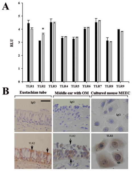
Figure 1.
Expression of the TLR profile in the human and mouse MEECs. Affymetrix microarray showed the expression of TLR1–TLR9 in the human NL-20 cells and TLR2 but not other TLRs significantly responded to the challenge of PGPS (gray bar) at 0.5 μg/mL for 8 h compared with PBS, black bar (A). Immunohistochemistry demonstrated that TLR2 was expressed in the human Eustachian tube epithelium (left, B, arrows pointing to the cellular membrane and the cytosol being full of mucous granules which are unstained and white in color), diseased middle ear mucosa (middle, B), and cultured mouse MEEC cells. Upper row in B, nonspecific IgG stain as controls for the lower row TLR2 stain; RLU, relative light unit; Scale bar = 20 μM applying to all panels in B; *p < 0.05, n = 3.

