Figure 2.
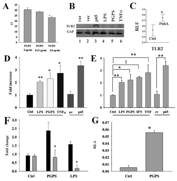
Pneumococci and their metabolites increased the expression of TLRs in vitro and in vivo. qPCR demonstrated that PGPS at 0.5 μg/mL significantly up-regulated the expression of TLR2 mRNA transcripts in mouse MEEC cells compared with control (A, p < 0.05, n = 3). Western blot analysis demonstrated that PGPS at 0.5 μg/mL, LPS at 0.1 μg/mL, TNF-α at 100 ng/mL, and p65 at 1.4 μg/mL up-regulated the expression of TLR2 at 6 h after recovery (B). Experimental OM verified that Pn6A significantly increased the expression level of TLR2 mRNA transcripts 3–14 d after bullar infection with Pneumococci (Pn6A) compared with those in controls using Affymetrix microarrays (C). ELISA demonstrated that bacterial products (PGPS and LPS) and inflammatory mediators (TNF-α and p65) at the same concentrations as above significantly up-regulated the expression of TLR2 protein in mouse MEEC (D, *p < 0.05, **p < 0.01, n = 3) and NL-20 human bronchial epithelial cells (E, *p < 0.05, **p < 0.01, §p < 0.001, n = 3). IκBαM (gray bar) significantly inhibited the TLR2 protein expression induced by PGPS and LPS compared with control, black bar (F, *p < 0.05, n = 3). PGPS at 1.25 μg/mL significantly increased the luciferase activity of TLR2 compared with its control PBS (G, p < 0.01, n = 9). ev, empty vector; Ctrl, control.

