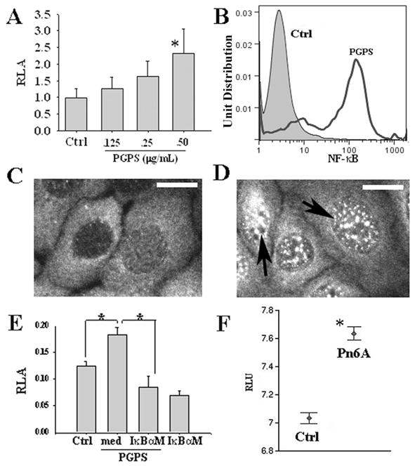
Figure 3.

PGPS induced the activity of NF-κB in mouse MEEC cells. PGPS at 0.5 μg/mL significantly increased the promoter activity of NF-κB compared with control (Ctrl) by luciferase assays (A, p < 0.05, n = 6). PGPS at 0.5 μg/mL significantly increased the percentage of activated NF-κB positive cells compared with PBS by FACS (B). PGPS incubation for 30 min increased the “activated” NF-κB translocation into the nuclei (D, black arrows) compared with control (C, bar = 5 μm) by immunohistochemistry. IκBαM significantly inhibited the promoter activity of NF-κB by PGPS at 0.5 μg/mL (E, *p < 0.05, n = 6). Note that IκBαM alone reduced the luciferase activity of NF-κB in nonPGPS treated cells. Pn6A infection in the middle ear of rats increased the expression of NF-κB mRNA transcripts 3–14 d after Pn6A infection in the middle ear of rats (F). *p < 0.05.
