
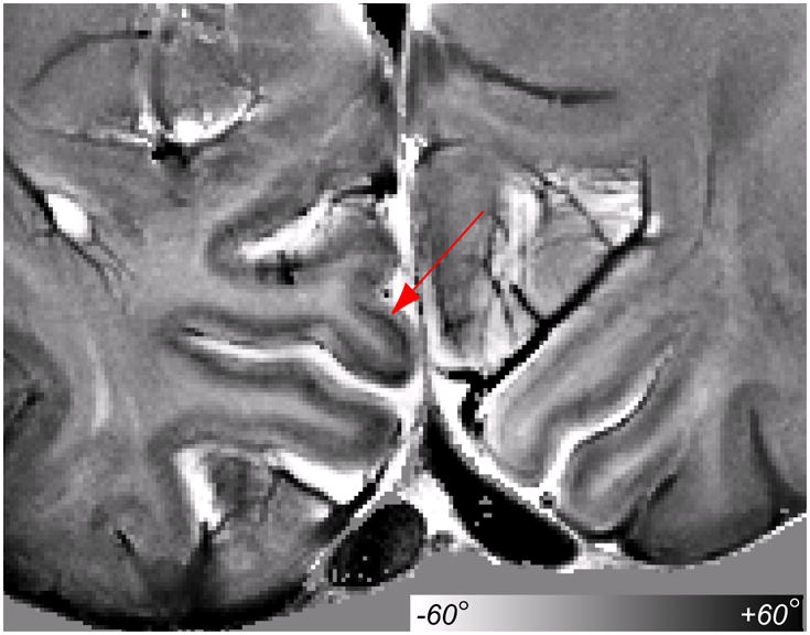
Figure 7.
High resolution bSSFP phase image at 3 T. The bSSFP method provides higher CNR that enables imaging of intracortical structure. The line of Gennari (red arrow) is visible at 3 T in vivo in a relatively short scan time (10 min).
Official websites use .gov
A
.gov website belongs to an official
government organization in the United States.
Secure .gov websites use HTTPS
A lock (
) or https:// means you've safely
connected to the .gov website. Share sensitive
information only on official, secure websites.
High resolution bSSFP phase image at 3 T. The bSSFP method provides higher CNR that enables imaging of intracortical structure. The line of Gennari (red arrow) is visible at 3 T in vivo in a relatively short scan time (10 min).