
Fig. 2.
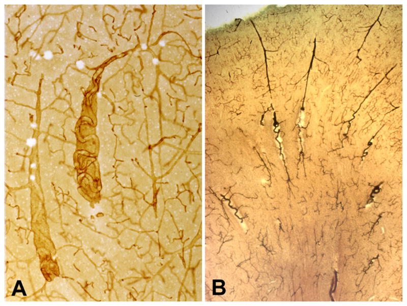
Tortuous arterioles in the white matter. (A) This thick celloidin section stained with collagen IV shows two tortuous arterioles in cavities. (B) This thick celloidin section stained with AP shows several arterioles with tortuosity beginning as they enter the white matter. (Reprinted from [20]).