
Fig. 3.
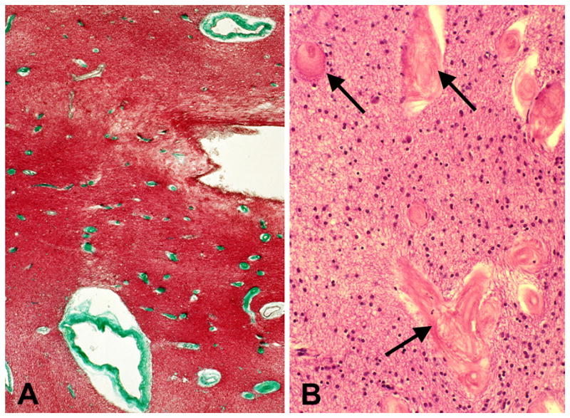
Severe periventricular venous collagenosis in the brain of a subject with leukoaraiosis. (A) This thin paraffin section stained with trichrome shows numerous affected veins (green) near the lateral ventricle. (B) This thin paraffin section stained with &E shows veins with collagenosis (arrows) at higher magnification. (Reprinted from [20]).