Figure 6.
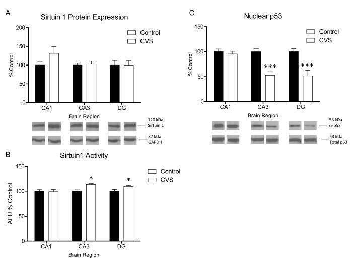
Regulation of Sirtuin 1 activity and protein expression in chronic stress. (A) Quantification and representative western blots of sirtuin 1 expression from nuclear protein extracted from subdissected hippocampus of control and chronic variable stress (CVS) animals. No significant difference from control animals in sirtuin 1 protein expression was seen in CA1, CA3, and DG regions in CVS animals (n=4). Representative blots from GAPDH are shown as protein loading control. (B) Quantification of Sirt 1 activity in nuclear extract from subdissected hippocampus of control and CVS animals. A significant increase in enzymatic activity of Sirt 1 was found in the CA3 and DG regions of the hippocampus of CVS animals compared to control animals. No change from control was seen in CA1 [data is represented as arbitrary florescence units (AFU) relative to control; n=4]. (C) Quantification and representative western blots of p53 acetylation relative to total p53 expression from nuclear protein extracted from subdissected hippocampus of control and CVS animals. A significant decrease in acetylation of p53 was observed in the CA3 and DG hippocampus regions of CVS animals compared to control animals while no difference from control was observed in CA1 (n=6). Data are shown as means ± SE; *P<0.05 vs. control; ***P<0.0001 vs. control.

